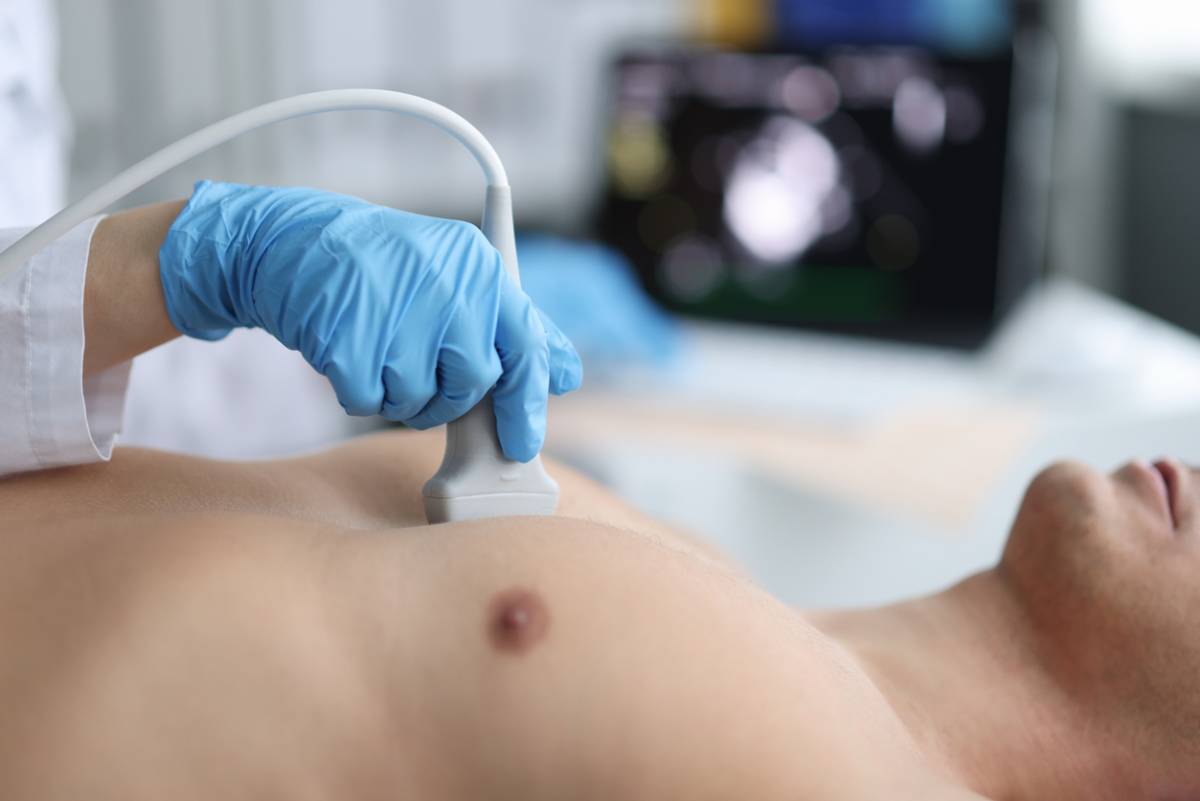
concept for which type of echocardiogram is best for you

Which Type of Echocardiogram Is Best for You?

Echocardiograms have long served as a staple in measuring heart health and function. What many patients do not know is that this is not a single test. Instead, several versions exist, each with its own functions. If you are worried about your heart health, you need to know which type of echocardiogram is best for you.

When you visit a heart health center for this exam, you will undergo one of three types of echocardiogram. These include:

  • Transthoracic echocardiogram
  • Transesophageal echocardiogram
  • Exercise stress echocardiogram

Which Type of Echocardiogram Is Best for You?

Heart imaging is a versatile practice that adapts based on the patient’s needs and suspected condition. In addition to different test types, the imaging technique can vary. Imaging modalities include:

Variations in Imaging Techniques

  • 2D Ultrasound: The most common approach, involves producing 2D images that can be stacked to render a 3D image of the heart.
  • 3D Ultrasound: A more advanced technique that evaluates the heart’s structures, blood-pumping capacity, and sounds.
    Doppler Ultrasound: Evaluates the pace and direction of blood flow.
  • Color Doppler Ultrasound: Similar to Doppler ultrasound, but uses color variety to highlight different vascular structures.
  • Stain Imaging: Demonstrates heart muscle movements, ideal for catching heart disease in early stages.
  • Contrast Imaging: Using an injected contrast agent, this can show better detail of the heart.

Transthoracic Echocardiogram

A transthoracic echocardiogram (TTE) relies on noninvasive ultrasound imaging (thus, the “echo” part of the name). It is the most common type and reliably provides imaging for all four of the heart’s chambers, the four valves, and surrounding blood vessel structures. By looking at factors like blood flow, chamber size, and blood pressure, it can check for the origins of concerning heart symptoms.

Who It Is Best for:

Patients with symptoms like:

  • Chest pain
  • Swelling
  • Heart murmurs
  • Dyspnea (shortness of breath)

Screening for conditions like:

  • Blood clots
  • Heart failure
  • Aortic aneurysm or dissection
  • Valve disease
  • Heart cancer
  • Hypertrophic cardiomyopathy

Transesophageal Echocardiogram

This method also uses ultrasound waves to generate imaging of the heart. However, it takes the pictures from within the body via an endoscopic device. Though slightly more invasive, it is typically well-tolerated. By taking imaging from within the body, it can create more detailed images of the heart and surrounding structures. It may use both Doppler and color Doppler techniques, depending on the information you need gathered.

Who It Is Best for:

Patients with a situation like:

  • Inconclusive results on a less invasive exam
  • Having a life-threatening issue that requires detailed imaging
  • Checking for blood clots before certain medical procedures
  • Confirming the results of certain procedures

Screening for conditions like:

  • Aortic aneurysm
  • Blood clots
  • Heart (cardiac) tumor
  • Congenital heart disease
  • Valve disease
  • Pericardial disease
  • Heart tissue/valve infections

Exercise Stress Echocardiogram

This unique type of echocardiogram involves ultrasound imaging taken before and after exercise. It looks for fluctuations in heart function when your cardiovascular system is placed under stress. It can involve a variety of techniques, depending on what your provider wants to screen for. It may also be used for monitoring known conditions to track improvements or disease progression.

Who It Is Best for:

Patients with a situation like:

  • Heart or vascular disease that requires monitoring
  • Showing symptoms of certain medical conditions
  • Athletes who need physical monitoring
  • About to undergo surgery

Screening for conditions like:

  • Cardiomyopathy
  • Congenital heart disease
  • Heart failure
  • Valve disease
  • High blood pressure in the lungs (pulmonary hypertension)

Which Do I Need?

The type of screening or exam you need must match your circumstances. When you meet with a cardiovascular specialist, they will determine screening modality based on your symptoms and medical history. Typically, imaging begins with the least invasive method. However, urgent situations may require immediately using the method best suited for high-quality images to diagnose and treat your condition.

Put Your Heart Health First

Visit your nearest heart health center as soon as possible if you have noticed troubling symptoms.

concept for common reasons for nipple discharge

Common Reasons for Nipple Discharge 

Nipple discharge is probably one of the most alarming things that can happen to a woman, but women should understand that it is relatively common and is not usually a sign of something serious.  However, it can also be an indication of different health conditions that may require attention at a medical center. It is normal for women to feel anxious upon noticing nipple discharge, but understanding the possible causes of nipple discharge can help you determine whether there is no real cause for anxiety or if you need to see a doctor immediately. Awareness about the common reasons for nipple discharge will help you get the right help if you need it.

Common Reasons for Nipple Discharge

Hormonal Fluctuations

Among the most common causes of nipple discharge is hormonal fluctuations. Women’s hormone levels fluctuate throughout their menstrual cycle, which can sometimes cause a small amount of milky fluid to leak out from the nipples. Although this discharge is not usually concerning, it is crucial for you to monitor the color and consistency and report any changes to the doctor.

Pregnancy and Breastfeeding

Pregnancy and breastfeeding are two other reasons women have nipple discharge. Pregnant women who are not yet breastfeeding may notice some discharge as their breasts prepare for milk production. On the other hand, women who have stopped breastfeeding may still observe a milky discharge months after stopping.

Fibrocystic Breast Changes

Fibrocystic breasts are characterized by lumps, which can be tender and swollen. Although this is a normal occurrence before a woman’s period arrives, it can lead to nipple discharge.

Infection or Abscess

Breastfeeding women can have mastitis or breast infection. This may result in nipple discharge. Although this is more common in breastfeeding women, it can also happen to anyone. The infection can cause a yellow or greenish pus-like discharge accompanied by breast pain, swelling, and fever. If the infection progresses, pus can build up. If you think you have an infection, visit your doctor immediately to prevent further complications.

Galactorrhea

This condition is experienced by women who are not pregnant nor breastfeeding, yet they produce milk. It is usually caused by hormonal imbalance, in particular, an elevation of prolactin, which is the hormone responsible for milk production. Other hormones that can affect milk production are estrogen and thyrotropin-releasing hormones. Thyroid disorders such as a benign tumor in the pituitary gland can cause this condition. In addition, medications like birth control pills or anti-depressants may also be responsible for galactorrhea. A physician can help you identify the problem and help address it.

Duct Ectasia

This is also known as mammary duct ectasia, which is a condition experienced by women nearing menopause. This does not mean, however, that women who are not yet menopausal do not have this condition. The fact is, it can happen to younger women too. This happens by a widening of a milk duct and thickening of its walls. This may cause a blockage to the milk duct, resulting in fluid build-up. If left untreated, this can lead to an infection.

Papilloma

These are small wart-like tumors that can cause clear or bloody discharge. Although they are usually harmless, a bloody discharge should not be taken for granted. This type of discharge may be a symptom of a more serious health condition like breast cancer. Due to this, it is crucial for you to visit a specialist for a thorough evaluation of the discharge. Remember that a timely diagnosis can greatly impact your prognosis.

Breast Cancer

As mentioned earlier, nipple discharge may be a symptom of breast cancer, especially if it is bloody or if it only comes out of one breast. A rare form of breast cancer called Paget’s disease of the breast can cause yellow or bloody discharge. Other symptoms of this cancer are itching, redness, an inverted nipple, a tingling or burning sensation, a lump in the breast, and thickening skin on the breast, among many others.

When to Visit Your Doctor?

Nipple discharge is usually not a sign of a serious health condition. However, every woman should observe their breasts for some signs that should prompt them to see their doctor.

  • Discharge that occurs even without any external interference like squeezing the nipple
  • Bloody or clear discharge only from one breast
  • If discharge is accompanied by a lump, redness, or breast pain
  • If a woman experiencing the discharge is postmenopausal

Your doctor will conduct a physical examination of your breast and possibly recommend that you undergo a digital mammogram for a thorough assessment.

Get the Right and Timely Treatment

There are many reasons for nipple discharge. Some are natural and harmless, while some need special attention from a licensed medical specialist. When it comes to your health, it is always wise to exercise vigilance. Monitoring your symptoms and visiting a healthcare specialist as soon as possible can help ease your worries and pave the way to early detection, treatment, and a more hopeful prognosis. If you cannot explain your nipple discharge, schedule a consultation with us today.

concept for how often should women have mammograms

How Often Should Women Have Mammograms?

A mammogram is an essential part of maintaining the long-term health of your breasts, as it remains the primary method for helping to identify and diagnose the presence of breast cancer and other conditions. Regularly receiving a mammogram should be considered a protocol for all women. But how often should women have mammograms?

How Often Should Women Have Mammograms?

However, the guidelines surrounding the frequency with which a woman should go in for a checkup can change, leaving many people confused about how often they should engage in this activity.

If you’re thinking it might be time for you to receive a mammogram, take a look at this guide that describes how often you should head in for a 3D digital breast mammogram.

What Is a Mammogram?

A mammogram is a type of diagnostic imaging test that is an X-ray of a woman’s breast. A mammogram is considered a first line of defense for helping a physician detect the presence of cancer and other conditions of the breasts.

Mammograms are considered highly safe and are completely painless as well. Following this, there’s no good reason for one to avoid engaging in this test on a regular basis. There is some advantage to returning to the same clinic and doctor for repeat tests, as this can help your medical professional study the health of your breasts over time.

How Often Should Women Have Mammograms?

Prior to the age of 40, most physicians recommend that a woman go in for a mammogram at least once every 3 years. After the age of 40, it is recommended that a woman receive a mammogram on an annual basis.

In addition, some doctors may recommend that a woman receive a mammogram annually, even before the age of 40. In addition, the frequency that you should receive a mammogram may increase if you have a history of conditions affecting your breasts, or if you have other types of health issues that might impact the health of your breasts.

What Occurs During a Mammogram?

During a mammogram, an imagist will compress the breasts between a set of firm surfaces to spread out the breast tissue and prepare it for imaging. An x-ray then subsequently takes a series of black and white images that are displayed on a monitor to be read by your technician.

At this point, your images will be forwarded to your general practitioner for reading and analyzing. If your imagist or physician feels that further testing is warranted, then you may be referred for additional procedures or to see a specialist of some kind.

If you are referred for additional testing, it’s not necessarily something to panic about. Mentally preparing yourself for the complicated reality of diagnostic imaging tests is a worthwhile activity when you’re engaging in these types of tests.

Are There Any Risks Involved?

Mammograms are considered very low-risk medical procedures. However, you should be prepared for the following elements of a mammogram test:

  • Radiation: A mammogram will expose you to a small amount of radiation.
  • Fallibility: A mammogram is considered a very accurate medical test. However, mammograms can’t detect all kinds of cancers. This is why the test should be paired with a physical exam.
  • Emotional Preparedness: Patients who receive a mammogram should be prepared to receive possibly extremely bad news if they are diagnosed with cancer. This includes the possibility of receiving a cancer diagnosis that is terminal, as many forms of breast cancer can be extremely aggressive.

Overall, the security provided by a mammogram is well worth the mild risks that are incurred by it. Preparing yourself emotionally in the case of disappointing results is a powerful step towards maintaining a state of health.

Preparing for a Mammogram

There are certain steps you can take to properly prepare yourself for a mammogram, as well as assist your medical personnel throughout the process. Some of the steps that can be taken in this regard include:

  • Bring any prior mammogram images and tests that you may have received.
  • Avoid wearing overly strong smells to your mammogram appointment
  • Schedule your test for a time when your breasts are likely to less tender, away from your ovulation

Your physician may have additional advice for your preparatory period, depending on your medical history. This will be gone over during a pre-screening consultation and preparatory period that will occur before the actual test.

The Best Imaging Clinic in Texas

West Texas Health is one of the most highly regarded imaging clinics in Texas. If you feel like you might be due for a diagnostic mammogram at our women’s health center, contact us to set up an appointment.

Featured image for How Are Nuclear Stress Tests Performed

How Are Nuclear Stress Tests Performed?

A nuclear stress test is a tool that helps diagnose heart issues. It is also highly effective in monitoring your heart health and assessing the effectiveness of specific treatment plans. The results of this test help your doctor understand how your heart is functioning, which is critical for them to identify issues and improve your heart health. You may wonder, how are nuclear stress tests performed? Below, you will find helpful information about what to expect before, during, and after the test.

How Are Nuclear Stress Tests Performed?

A nuclear stress test is helpful in learning more about how your heart is working. The test involves two parts. If you have been scheduled for a nuclear stress test, it is important to consult with your doctor about how to prepare. They will provide you with helpful information about preparation, what to expect from their specific treatment process, and what will happen after. Below are general guidelines for how nuclear stress tests are performed.

Before the Nuclear Stress Test

You will need to physically prepare before your nuclear stress test. Things patients are most often told to avoid include eating, drinking, or taking certain medications for some time before your test. Your doctor will provide you with specific information about this. It is essential to follow their instructions carefully. Do not stop taking any medications without consulting your doctor first, as this can be dangerous and may impact your results. On the day of the exam, wear comfortable clothes. You should be able to exercise in them if the test calls for it.

During the Nuclear Stress Test

A nuclear stress test is performed at a cardiovascular health center. The nuclear stress test starts with an injection. The injection is a small amount of radioactive material. It is referred to as a tracer and is injected into your arm. The injection travels through your bloodstream to reach your heart. It is then absorbed by your heart. A special camera will then take a set of images while you are lying down. They show how your blood flows to your heart when you are resting. 

The second phase of the test involves assessing your heart when your body is not at rest. This portion of the exam is completed by measuring your heart during exercise or with medication. During this time, another set of images is taken. This shows the doctor how well your blood flows to your heart when it is under stress. Throughout the entire exam, several things are monitored, including your heart rate and blood pressure.

After the Nuclear Stress Test

Once the test is complete, you will be asked to rest for a short period. The doctor will observe you to ensure that your heart rate normalizes. The doctor will assess the images and review them with you. In some cases, you may need to set up a follow-up appointment to discuss your results. Your doctor will let you know what to expect after the nuclear stress test in advance. 

Do I Need A Nuclear Stress Test?

Your nuclear cardiology doctor will determine if you need a nuclear stress test. There are several reasons why you may need a nuclear stress test. The most common reasons are as follows.

Diagnosing Heart Conditions

A nuclear stress test assesses whether someone has coronary artery disease. It is also helpful in diagnosing several other heart conditions and assessing the severity of heart disease.

Treatment Assessment

If you are undergoing treatment for heart issues, a nuclear stress test can help assess your treatment. Your doctor can use the information to determine whether other treatments are necessary or if you are on the right track.

Apex Cardiology

The team of experts at Apex Cardiology is skilled in nuclear stress tests. If you feel you need to schedule an appointment for a nuclear stress test, contact their team today! 

A woman sitting alone with a bandage over her eye as the featured image for Orbital Trauma Treatment Options

Orbital Trauma Treatment Options

Orbital trauma refers to a spectrum of physical injuries, diseases, and congenital disorders that hurt the orbital area. Physical injuries and lesions on the eyelids may require orbital trauma care. Conditions such as ptosis and blepharospasms can worsen overall eye health when left untouched by surgical intervention. Depending on your individual needs, we will recommend the best avenue of treatment. 

What treatments are available for orbital trauma at the office of our oculofacial reconstructive surgeon? This quick guide will discuss specific forms of orbital trauma and their most effective treatment options. 

What is Orbital Trauma?

The area around your eyes is susceptible to damage. A blow to the eye can cause a myriad of orbital issues, leading to physical and medical trauma. A car accident or a collision with a wall, for example, can cause a variety of problems around the eyes. 

The most common type of orbital trauma is an orbital fracture. Orbital fractures occur when the fragile bony structure surrounding your eye is hit with such a force that it breaks. Such impact can cause a fracture that “blows out” your orbital bone and causes it to fall out of alignment. While some minor cases of orbital trauma may not need surgical intervention, anyone dealing with symptoms of pain and swelling due to an orbital fracture should receive treatment right away.

Orbital Trauma Treatment Options

The first step to determine what course of treatment is best for you is to schedule a consultation with our surgeon. During your appointment, give as many details as possible about the type of injury you are dealing with. Aside from a bony fracture, orbital trauma can also indicate underlying issues such as ptosis or Graves’ Disease. The doctor will want to rule out as many causes as possible to find the best solution. 

At the office of Dr. Jennifer Murdock, we typically offer three distinct treatments for orbital trauma: 

Reconstruction

Full reconstruction is usually only used as a last resort, necessary only in cases of extreme emergency trauma. During reconstructive surgery, the surgeon uses various techniques, such as bone grafting and mesh reconstruction, to create a stable orbital structure. 

Reconstructive surgery can also involve orbital decompression. Certain conditions, such as emphysema, can put undue pressure on the orbital area, causing significant pain and swelling behind the eye. Orbital decompression releases some of this pressure, creating a larger space for your eyes to rest in their proper place. 

Orbital Implants

You might have gone through an injury or illness that has caused your eye to be removed. If you are missing an eye, you can receive an orbital implant in your eye sockets. This natural-looking implant can support the structure of your orbital bone while also providing great aesthetic appeal. 

Orbital implants are a recommended course of action for anyone struggling to regain confidence after losing an eye. We can carefully create and place a custom implant that will improve your facial structure and give you confidence the next time you look in the mirror.

Fracture Repair

A more common type of orbital trauma surgery that we provide includes fracture repair. Fractures can range from mild to severe, so it is important to check with your doctor first to see if surgery is the correct way to go. Major orbital fractures happen when part of the orbital bone breaks away from the rest of it, creating a spatial gap. Orbital surgery can use techniques like skin and bone grafting to create a support around the broken piece of bone.

Reconstruction from Previous Surgery

Orbital surgery requires a high level of precision and expertise. From time to time, a surgeon can make a mistake, leaving you with less-than-favorable results. Your botched surgery might also result from an injury that occurred during the healing process. Whatever the reason for your current dissatisfaction with the state of your orbital bone, you can opt for reconstructive surgery. 

Find Treatment for Your Orbital Trauma Today

Whether your orbital trauma is new or has been building up over a long period, appropriate treatments are available to fit your specific needs. Get informed about orbital trauma and the different types of injuries and conditions that might warrant the need for surgery. 

Visit Dr. Jennifer Murdock, a top-rated oculofacial reconstructive surgeon in Miami, to schedule your consultation. Orbital trauma only worsens the longer it is left untreated, so it is a good idea to act fast. Get both reconstructive and cosmetic repair to leave you feeling refreshed, healthy, and whole again. 

Featured image for When Should You Go For Cardiac CT Imaging

When Should You Go For Cardiac CT Imaging?

A CT scan is a medical imaging tool used to see the heart and blood vessels. The results allow your doctor to see any potential issues in your heart. It can also be used to monitor your progress if you are undergoing medical treatment for your heart. The procedure is non-invasive and highly effective. Below, you will find information detailing when you should go for cardiac CT imaging.

When Should You Go For Cardiac CT Imaging?

A CT scan is also referred to as a cardiac computed tomography scan. It is highly effective in detecting several heart conditions because it examines your heart and blood vessels. Your primary care provider or cardiology doctor may recommend it. It is critical to follow their instructions, as the results can provide vital information about your health. There are several reasons someone should get a CT scan.

Coronary Artery Disease

CT scans are helpful in detecting coronary artery disease. The imaging detects blockages and narrowing of the arteries, among other important information. This information can reduce your risk for heart attacks and other cardiac complications. Your cardiology doctor uses the information to create an effective coronary artery disease treatment plan.

Congenital Heart Defects

Congenital heart defects can impact your heart and overall health. A CT scan effectively gets images of the entire heart. These details allow a cardiology doctor to diagnose and monitor the defect. Undiagnosed congenital heart defects can result in serious health issues. Medical intervention is key, and a CT scan can help your doctor create a treatment plan that is right for you. 

Heart Valve Issues

Heart valve issues can impact your health and lead to serious health issues. Valve issues often involve narrowing or leakage. A CT scan can evaluate the function of your heart valves. This information can aid your cardiology doctor in finding a treatment plan for you. Surgical intervention may be necessary. This is why it is critical to get a CT scan when your doctor recommends it.

Management and Evaluation

CT scans are important when diagnosing several heart conditions. However, they are also an effective tool for evaluating and monitoring heart conditions. They can assess how treatment is going for a heart condition and be used before or after surgery to see any changes that have occurred. CT scans are a highly valuable tool used by cardiology doctors to ensure your heart health stays in good shape.

Non-Invasive Cardiac CT Imaging

Preparing for a CT scan is simple. It’s helpful to follow your cardiology doctor’s instructions. This may include fasting, adjusting your medications, and following other pre-scan instructions. You should also avoid wearing jewelry when you go to your appointment. Be sure to consult with your primary care provider before adjusting any of your medications. 

The scan is non-invasive and fairly fast. You should be in and out of the office within 90 minutes. Non-invasive cardiac CT imaging involves several steps. You will need to lie down on a table during the scan. Electrodes may be placed on your chest to monitor your heart rate. A dye is injected into your body to improve the clarity of the CT scan images. From there, you need to stay still and may be prompted to hold your breath. Once the imaging is complete, a radiologist will analyze the images. From there, your doctor will reach out to you regarding your results. 

Apex Cardiology

The team at Apex Cardiology has helped countless patients improve their heart health. If you need a CT scan, contact Apex Cardiology to schedule your appointment!

Illustration of woman of different ethnicities in solidarity to represent benefits of a regular breast examination

5 Benefits of a Regular Breast Examination

Breast examinations can cause a woman anxiety for several different reasons. However, there are many benefits to a regular breast examination. In fact, regular breast checkups are an essential component of maintaining optimal breast health during a woman’s lifetime. Failing to engage in this essential process can leave an individual unaware of emerging health risks.

Knowing the ways that this can benefit you can encourage people to more consistently engage in this process, which ultimately has the potential to ensure better health in the long run. If you are on the fence about having a breast examination, take a look at this overview of the ways that an in-office mammogram can help you.

What Occurs During a Breast Examination?

During a breast examination, your physician will examine your breasts to determine whether or not there are abnormalities present. Should they determine that there is something to investigate further, then they will likely refer you for further testing and imaging work.

Overall, the entire process takes little time. It is usually completed within minutes. This allows patients to complete their examination at a convenient time of their choosing.

Although receiving a breast examination can be intimidating or anxiety-provoking, it beats the alternative, as failing to engage in this essential health practice can leave one completely unaware of the internal health of their breasts.

5 Benefits of a Regular Breast Examination

1. It Can Give You Peace of Mind

One of the main benefits of consistent breast examinations is that they have the potential to offer a patient peace of mind. Regardless of whether you ultimately have an issue with your breasts or not, getting them consistently examined can give you a clear answer and dispense any ambiguity.

Knowledge is power, and this applies to one’s health as well. Taking control of your relationship with your body can leave you feeling more empowered and able to respond to the potential issues that arise.

2. It Can Alert You to Developing Problems

One of the main advantages of receiving consistent examinations is that it is the most surefire way to alert you to potential developing health issues in your breasts. For almost all major health issues, it pays to catch issues that are developing early, as this increases the likelihood that you will be able to solve the issue with less stress.

In addition, taking care of issues on the earlier side can prevent further complications with other aspects of your health. Overall, it

3. It Can Give Your Doctor a Portrait of Your Health History

Medical professionals are able to offer individuals a higher level of medical care if they have a clearer portrait of an individual’s health history. If you stick to receiving treatment from the same technician, then there is a higher chance that you will be able to build a more intimate relationship with them in terms of how they understand your progress or lack of progress in major areas of your health.

This can often mean the difference between receiving high-quality care from someone who understands your body and being rushed through a process with a doctor who does not take your needs into account.

4. It Can Possibly Save Your Life

Although relatively rare, breasts can face very serious ailments, such as breast cancer. Failing to catch these conditions as they develop can have dire consequences, as it is essential to catch these types of diseases early in order to lessen their impact.

If you catch conditions like breast cancer early, you will have a significantly higher chance of beating it and maintaining a higher standard of health throughout your life. On the other hand, failing to catch cancer at the appropriate time can have deadly consequences.

5. It Can Help You in Other Medical Areas

If you determine whether or not a problem you are dealing with originates in the breasts, it can help you rule out certain health complications. This can help you determine what the correct path forward is for taking the best care of your health. Overall, as a quick and effective method of diagnosis, there’s no reason not to engage.

Your Resource for Breast Examination in Abilene 

West Texas Health is one of the most trusted clinics in Texas for providing patients with high-quality examinations and imaging care. Now that you know the benefits of a regular breast examination, contact our clinic today to discuss your options for moving forward with one in Abilene.

Should You Get Nasolacrimal Surgery?

Nasolacrimal surgery is a procedure that helps patients with tear duct blockage. If you notice that you cannot properly cry or your eyes are constantly watering without leaking, you might be dealing with a blocked duct. There are a few reasons why you might be experiencing nasolacrimal obstruction. It is essential to identify what is causing this unhealthy buildup. After speaking with our surgeon, you will know if you should get nasolacrimal surgery.

Nasolacrimal surgery should be performed by a reconstructive plastic surgeon who has plenty of experience with this procedure. Learn more about the common symptoms and causes of tear duct blockage and find out what nasolacrimal surgery entails by reading the article below. 

Causes of Tear Duct Obstruction

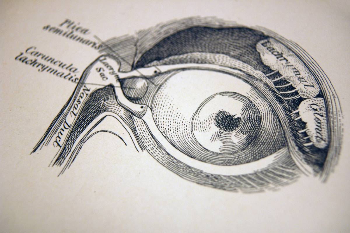
Your nasolacrimal duct is responsible for the release of tears, which drain toxins and other debris from your eye. Tears are necessary if you want to keep your eyes and ducts as healthy as you can. While this does not mean you need to start crying every day, it can be a cause for concern if you cannot cry at all. 

Your tear ducts might be obstructed for a number of reasons. First, you might be born with a congenital condition. It is common for babies to be born with blocked tear ducts, for example. This usually is not a cause for concern since the tear ducts become naturally unblocked within a year or so. However, you might have also had a physical injury or trauma that damaged this part of your face. Certain ailments and even some medical treatments can also cause the tear ducts to get blocked. 

Symptoms of Nasolacrimal Blockage

If you are struggling with tear duct blockage, you should go down this list of symptoms to see if your case is so severe that it warrants nasolacrimal surgery. There is a difference between finding it difficult to cry and having a real blockage that allows debris to build up in your tear ducts. 

Some of the most common symptoms of a blocked tear duct include: 

  • Redness and puffiness around the eyes, especially the lower eyelids
  • Your eyes are constantly tearing up.
  • Your vision gets blurry.
  • There is mucous coming from your eyes.
  • You are consistently swollen or irritated around the eyes.
  • You cannot properly blink.
  • You get eye infections often, including conjunctivitis (pinkeye) 

If you are experiencing these symptoms, contact your doctor immediately. Depending on how long you have dealt with these issues, you might need urgent treatment. 

What Nasolacrimal Surgery Entails 

The doctor will perform a series of tests to properly diagnose you with blocked nasolacrimal ducts. This usually includes eye irrigation, vision tests, and other types of imaging. After determining the extent of your blockage, a treatment plan will be suggested. Antibiotic eye drops, and rest can sometimes relieve the blockage. In other cases, nasolacrimal surgery might be necessary. 

Dacryocystorhinostomy

Tear duct surgery, also called DCR, works in one of two ways. It can either repair a blocked tear duct or create an alternate pathway for tears to flow down, essentially solving the blockage problem. You can speak with your doctor about whether or not you want to bypass your current blockage or get rid of it completely. 

This surgical procedure requires anesthesia during surgery. Your surgeon may use external techniques or may need the assistance of a tube that goes up your nose during the procedure. Depending on the severity of your needs, you can avoid visible scar tissue as soon as you are healed. 

Recovery Time

Once you wake up from your procedure, you will feel a little uncomfortable. There may be gauze in your nose to prevent bleeding and other bandages around your face. You should be able to go home on the same day. Make sure you take your prescribed antibiotics and eyedrops home with you, as you should start with them right away. 

As long as you follow your doctor’s instructions following surgery, you should have a quick and easy recovery. Most of our patients heal up within a few weeks, boasting beautiful results and enjoying your newly repaired nasolacrimal ducts. 

Fix Your Tear Ducts with Nasolacrimal Surgery Today

The symptoms of a blocked tear duct can be frustrating and painful. If you are tired of consistently swelling and tearing up, it might be time to unblock your tear duct with our innovative surgical procedures. Visit Dr. Jennifer Murdock’s office to learn more about tear duct blockage and the appropriate treatment plan for you. 

The image shows a man during an echocardiogram to show who is a candidate for echocardiography.

Who Is a Candidate for Echocardiography?

An echocardiogram is a vital medical test. Often referred to as an “echo,” the test is essentially an ultrasound of your heart. Doctors use the test to look for issues with your heart. It tests how blood moves through the chambers and valves of your heart. Your primary care provider or cardiologist can order an echocardiogram if they are concerned about your heart health. If you have any concerns about your cardiovascular health, you should reach out to your doctor. In the meantime, this guide will explain who is a candidate for echocardiography and if you fall into this category.

Who Is a Candidate for Echocardiography?

There are several reasons someone may need an echocardiogram. The text examines your heart and can determine abnormalities. Consider the following reasons why someone may be a candidate for an echocardiogram.

Congenital Heart Disease

Some people are born with heart defects, which are characterized by problems with the formation of the heart. An echocardiogram is effective in assessing formation issues, such as a hole or an abnormal connection. Untreated heart defects can cause serious issues later on.

Heart Murmurs

Unusual sounds from your heart characterize a heart murmur. Doctors hear them when they use a stethoscope to listen to your heart. Murmurs occur when blood abnormally flows through your heart. An echocardiogram is used to see what is happening inside your heart.  

Heart Valve Issues

Your heart has four valves. The valves open and close to let blood flow in the right direction. Sometimes, valves may become too right or leak, which can cause health issues if left unaddressed. An echocardiogram provides your cardiology doctor with helpful information to treat the heart valve issue.

Blood Clots

Blood clots can form inside your heart. If left unaddressed, they can be highly dangerous and fatal. Echocardiography in Los Angeles is often used to determine if a patient has a blood clot. This is essential information for your doctor, as they can use it to treat the issue. 

Symptom Evaluation

Some patients have several symptoms that indicate heart issues. Common symptoms include chest pain, dizziness, shortness of breath, and a fast or slow heartbeat. An echocardiogram is beneficial, as your doctor can see if the symptoms are associated with your heart. From there, they can diagnose and treat the issue much more easily.

Echocardiogram Testing

An echocardiogram typically takes roughly 30-60 minutes from start to finish. You will be on a special bed in a hospital gown, so the equipment can easily be placed on your chest area. The technician will then apply gel to your chest so they can produce a more clear image. A special tool called a transducer is used to take the echocardiogram. The technician will move the transducer around your chest to get as many quality images as possible. Once the technician has taken all of the information they need, the test is over. From there, the doctor will examine your results and get back to you regarding what they found.

Echocardiography in Los Angeles

If you are at high risk for developing heart conditions, it is crucial to work with a skilled cardiologist. They can assess your risk for certain conditions and provide you with important information about how to care for your health. If you are not high-risk but have concerns about your health, consult with a medical professional right away. The team at Apex Cardiology is skilled in cardiology and has provided countless echocardiograms. Contact their team today to schedule an appointment! 

The image shows a sign with "Pollen Season Ahead" to introduce steps you can take to prepare for seasonal allergies.

5 Steps You Can Take to Prepare for Seasonal Allergies

Allergies can affect people year-round, contributing to a series of negative symptoms and traits. You might be affected by the pollens that fly through the air during the spring. Or perhaps you are allergic to grass and deal with itching and irritation during the summertime. No matter what time of the year you deal with seasonal symptoms, you should invest in professional allergy services and take steps to prepare for seasonal allergies. 

If you feel allergy symptoms are starting to creep up on you, read on to learn steps you can take to prepare for seasonal allergies.

5 Steps You Can Take to Prepare for Seasonal Allergies

1. Learn Your Allergy Triggers

Different allergies stem from different triggers and allergens. It is important to recognize what exactly sparks your allergic reactions. For example, if you are allergic to flower pollens, stay indoors on particularly windy days. If you have multiple allergic reactions and cannot quite pinpoint the source, we encourage you to visit us for a comprehensive allergy test. 

2. Learn When Allergy Season Starts

Season allergies can occur every few months. Those with severe reactions might feel like they are always coming down with different allergy symptoms. Knowing when to expect seasonal flare-ups can help you prepare for the difficult times ahead. More importantly, knowing what to expect can help you avoid these problems altogether. 

Most people experience spring allergies, which can begin as early as February and last through the summer. Late summer and fall allergies exist as well, as the changing leaves and intense weather patterns take a toll on our bodies. Avoiding pollens, mold, dander, and other possible allergens during these peak times will keep your symptoms at bay. 

3. Equip Your Home with the Essentials

During particularly bad bouts of weather, you may want to stay indoors and avoid an allergic reaction—let alone a cold. If you are already struggling with an outbreak of allergies, these at-home essentials can help: 

  • Combine your air conditioner and dehumidifier to create a cool and dry environment, free of moisture in the air. 
  • Use designated filters and HEPA items to keep even the most microscopic of allergens out of the air in your home. 
  • Try to sweep and vacuum often to keep the area clean. 

If your allergies are very serious, you might also be prescribed medication like an inhaler or an EpiPen for use in an emergency.

4. List and Track Your Symptoms

If you are ready to get medical help for your seasonal allergy problems, you have come to the right place. When you reach out to our West Texas Health experts, we will ask you to describe the exact symptoms you are experiencing. Depending on the type of seasonal allergies you get, as well as how intense your symptoms are, there might be a specific type of treatment that works for you. 

Many allergy symptoms are similar to those of a common cold. Fever, sore throat, runny nose, congestion, coughing, watery eyes, itchiness, rash, and fatigue can all be signs that you are having an allergic reaction. 

5. Get Started with an Appropriate Treatment Plan

The longer you go without seeking appropriate care for seasonal allergies, the worse your symptoms can get. Instead of waiting for your health to deteriorate, you can get essential medical care in Abilene by reaching out to our friendly experts today. We can help you narrow down your allergy symptoms to pinpoint the exact trigger. With a combination of education and prescribed medication, you can set yourself up for an allergy-free future. Get started with us today to learn more!