Preparing for Your Echocardiogram
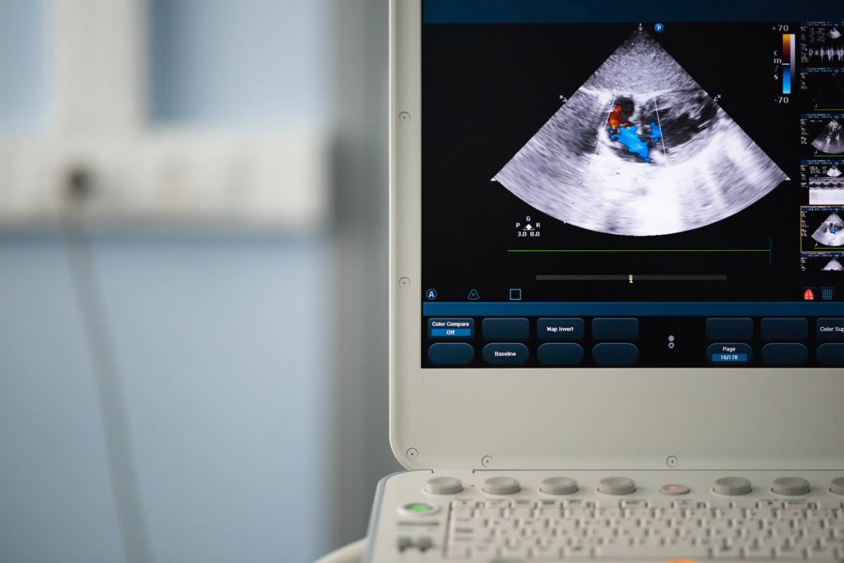
An echocardiogram is a type of sonogram that uses sound waves to create an image of the heart’s function. In this way, echocardiograms are life-saving, medical marvels. This type of sonogram allows doctors to recognize how the heart is beating and pumping blood.
Using echocardiographic technology, doctors can better diagnose and treat patients with cardiovascular or heart-related conditions. Echocardiogram providers can assess the severity of your condition by examining the heart closely with an echocardiogram.
What Patients Should Know Before an Echocardiogram
Before receiving an echocardiogram, patients should know that there are a multitude of echocardiogram tests. Depending on your cardiovascular health and symptoms you exhibit, doctors will determine which echocardiogram test is best for you to undergo.
In addition, patients should understand that receiving an echocardiogram is helpful for early intervention. When caught early, doctors can help reduce the effects of cardiovascular issues. This can prove to be life-saving.
How to Prepare for Your Echocardiogram
Although you may feel nervous or anxious about your echocardiogram test, understanding how echocardiograms work will put you at ease. While tests may differ, each focuses on the heart, detecting any cardiovascular problems along the way.
To prepare for an echocardiogram, consider:
- Providing a list of symptoms you experience to your doctor
- Following your doctor’s instructions for eating and drinking before your procedure
- Arranging a ride home if the procedure requires sedation
- Avoiding wearing jewelry such as necklaces or rings
- Wearing loose-fitting or comfortable clothes
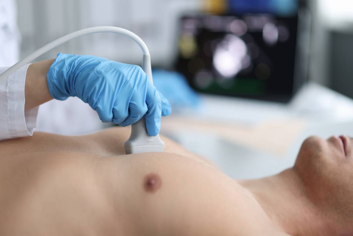
According to the National Library of Medicine, preparations are minimal. On the day of the procedure, once you arrive at the designated location, you will be asked to remove clothing covering your chest. You may also receive cardiac monitoring stickers so doctors can monitor your heart.
Types of Echocardiogram Tests
Not all cardiovascular issues are the same. Heart-related problems vary from person to person. Thankfully, echocardiograms are available for a number of tests.
In fact, the following tests are offered to candidates:
- Stress echocardiogram – A type of test performed after the heart is stressed. This typically involves the patient performing exercises or receiving a medication to make the heart beat more rapidly. This echocardiogram can help patients learn if they have issues with blood flow or coronary artery disease.
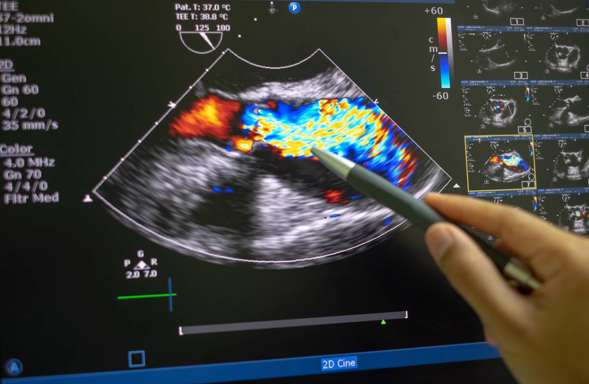
- Doppler echocardiogram – A type of test examining the way blood flows through the heart, blood vessels, and heart valves.
- Transesophageal echocardiogram – This type of test involves a probe passing down the esophagus instead of moving across the outside of the chest. As a result, patients receive higher-resolution photos of the heart.
- Transthoracic echocardiogram – This type of echocardiogram procedure uses the heart rate obtained by moving the transducer to different locations on your chest.
The type of test your doctor assigns to you will depend on your condition and the cardiovascular symptoms, if any, you experience.
Why Echocardiograms Work
Echocardiograms are a non-invasive method for examining cardiac anatomy. Although there are different types of echocardiograms, the echocardiographic process provides thin cross-sections of cardiac structures, including the left and right atria, the left and right ventricles, and any other structures associated with heart valves.
Assessing cardiac muscles during contractions is a form of stress test. More invasive procedures may be needed if patients score poorly on the stress test. Thankfully, echocardiograms allow doctors to visualize the heart chambers and detect potential health concerns.
Schedule an Echocardiogram
At Apex Cardiology, our cardiologists provide high-quality care. We focus on every activity and treatment to offer quality care.
We have a long history of treating heart conditions. Whether you struggle with stress or an underlying condition, our team of experts can provide you with the healthcare you deserve.
For those experiencing cardiovascular issues, contact us today to speak with our cardiologists and learn about your treatment options.