Autoimmune Diseases That Affect Vascular Health

Autoimmune diseases occur when the immune system mistakenly attacks the body’s own tissues, and when blood vessels are targeted, serious complications can develop. If you are experiencing these complications, you should often seek care at a vascular treatment center, where specialists focus exclusively on preserving vascular health and preventing organ damage. These conditions can cause symptoms such as inflammation of arteries, veins, and capillaries throughout your body. This can lead to reduced blood flow, clot formation, and tissue trauma. You can be proactive about your vascular health by recognizing early warning signs and staying up to date on the latest treatment options.

Autoimmune Diseases and Vascular Health

When your immune system attacks blood vessels, it triggers inflammation that damages vessel walls and disrupts circulation. This inflammation can affect arteries, veins, and capillaries throughout the body, compromising vascular health and often going unnoticed until damage occurs. Timely intervention of specialized vascular treatment when autoimmune diseases and vascular health collide can prevent irreversible damage and preserve quality of life and longevity.

Long-term Complications of Chronic Vascular Inflammation

Persistent inflammation progressively weakens blood vessel walls, leading to a variety of complications. When left untreated, reduced blood flow can create organ damage. Chronic inflammation can also accelerate the progression of several life-threatening conditions. Many complications are cumulative and often irreversible once established. Early aggressive treatment is essential. Regular monitoring at a vascular treatment center helps detect the silent progression before catastrophic events occur.

Symptoms and Warning Signs

Autoimmune vascular disease presents with symptoms that are both localized and systemic. Persistent fatigue, unexplained fever, weight loss, and night sweats may be indicators that something needs further attention. Vascular-specific warning signs vary depending on the affected vessels. Talk to your specialist if you experience:

  • Discoloration of the fingers
  • Painful skin ulcers that won’t heal
  • Sudden numbness or weakness in limbs
  • Visual disturbances
  • Abdominal pain
  • Chest pain, headaches, jaw pain, or shoulder tenderness

Any combination of these symptoms should prompt immediate medical attention for professional evaluation to prevent permanent damage.

Specific Autoimmune Diseases that Affect Vascular Health

It is important to monitor symptoms and take the steps necessary to reduce your risk of vascular complications related to autoimmune diseases. Autoimmune diseases that particularly affect vascular health include:

  • Systemic Lupus Erythematosus
  • Rheumatoid Arthritis
  • Systemic Scleroderma
  • Antiphospholipid Syndrome
  • Giant Cell Arteries
  • Behcet’s Disease
  • ANCA-associated vasculitides
  • Sjogren’s Syndrome
  • Polyarteritis Nodosa

Lifestyle Management and Risk Reduction

Managing autoimmune vascular disease requires conscientious lifestyle modifications. Quitting smoking is essential, and following a diet rich in omega-3s, fruits, and vegetables while limiting processed foods is a good start in reducing the risk of complications. Moderate exercise improves circulation, and stress management is beneficial as well. Most importantly, maintain strict adherence to medication instructions and attend all follow-up appointments to catch complications early.

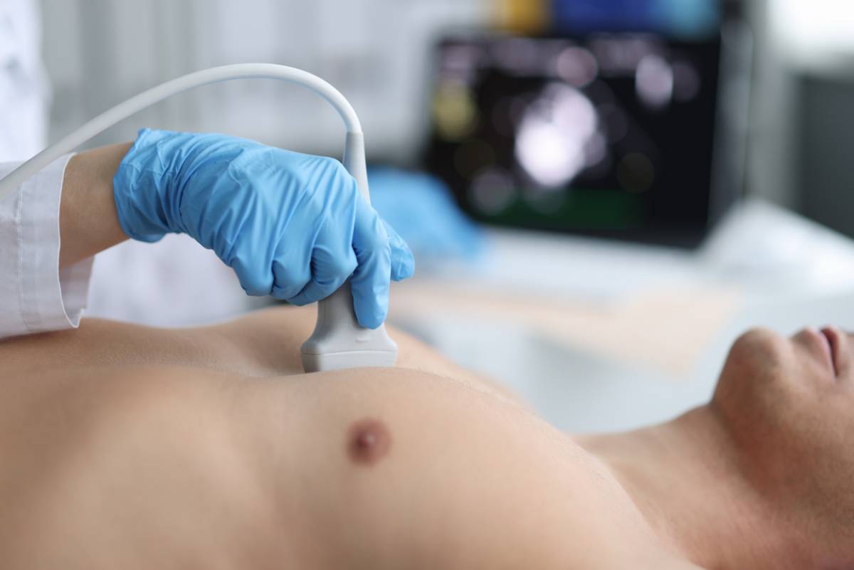
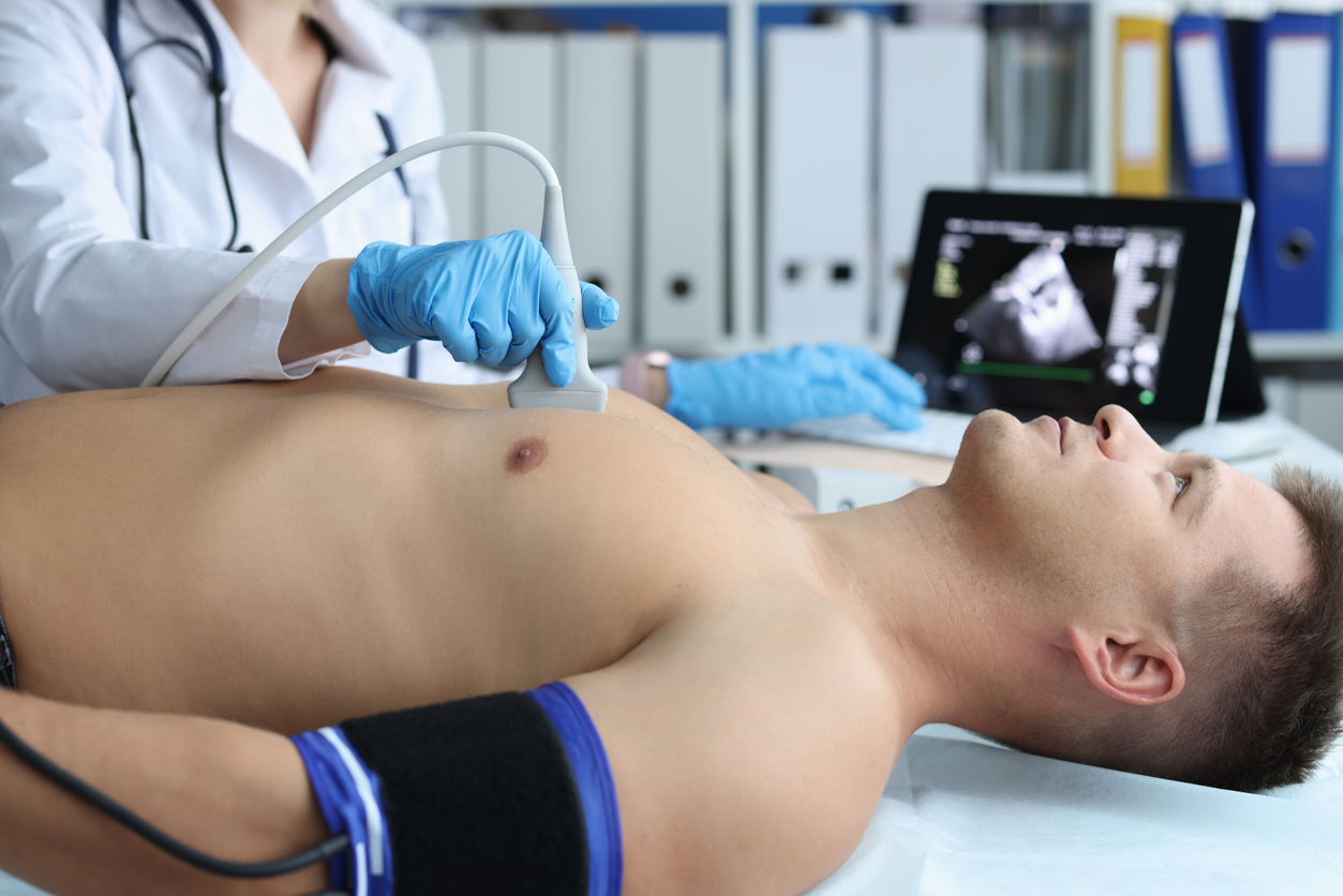
Diagnosis and Treatment

Blood tests indicating elevated inflammatory markers and autoantibodies are the initial step in diagnosis. Imaging studies are used to visualize vascular inflammation and track blood flow. When imaging proves inconclusive, a biopsy of the affected tissue is taken and tested to confirm the diagnosis. Coordinated care among medical professionals ensures that both the autoimmune disease and any other conditions that may complicate it receive targeted, effective management for optimal outcomes.

Protecting Vascular Health from Autoimmune Diseases

Autoimmune diseases that target vascular health demand extra vigilance and specialized care to effectively prevent devastating complications. The connection between immune system dysfunction and vessel damage can have devastating, sometimes fatal consequences when not adequately addressed. These conditions are serious,s and modern diagnostics and targeted therapies offer excellent control when managed by experienced specialists. Regular monitoring of vascular health, combined with diligent self-care and careful adherence to all instructions provided by your experienced care team, can significantly improve long-term outcomes. By prioritizing early intervention and maintaining consistent follow-up, you can effectively manage your condition, preserve organ function, and maintain quality of life despite autoimmune-driven vascular challenges.

Don’t Wait

Schedule your consultation at our vascular treatment center today to discuss your symptoms and develop a personalized treatment plan.

Doctor using stethoscope for patient examination.

Preparing for Your Echocardiogram

An echocardiogram is a type of sonogram that uses sound waves to create an image of the heart’s function. In this way, echocardiograms are life-saving, medical marvels. This type of sonogram allows doctors to recognize how the heart is beating and pumping blood.

Using echocardiographic technology, doctors can better diagnose and treat patients with cardiovascular or heart-related conditions. Echocardiogram providers can assess the severity of your condition by examining the heart closely with an echocardiogram.

What Patients Should Know Before an Echocardiogram

Before receiving an echocardiogram, patients should know that there are a multitude of echocardiogram tests. Depending on your cardiovascular health and symptoms you exhibit, doctors will determine which echocardiogram test is best for you to undergo.

In addition, patients should understand that receiving an echocardiogram is helpful for early intervention. When caught early, doctors can help reduce the effects of cardiovascular issues. This can prove to be life-saving.

How to Prepare for Your Echocardiogram

Although you may feel nervous or anxious about your echocardiogram test, understanding how echocardiograms work will put you at ease. While tests may differ, each focuses on the heart, detecting any cardiovascular problems along the way.

To prepare for an echocardiogram, consider:

  • Providing a list of symptoms you experience to your doctor
  • Following your doctor’s instructions for eating and drinking before your procedure
  • Arranging a ride home if the procedure requires sedation
  • Avoiding wearing jewelry such as necklaces or rings
  • Wearing loose-fitting or comfortable clothes

According to the National Library of Medicine, preparations are minimal. On the day of the procedure, once you arrive at the designated location, you will be asked to remove clothing covering your chest. You may also receive cardiac monitoring stickers so doctors can monitor your heart.

Types of Echocardiogram Tests

Not all cardiovascular issues are the same. Heart-related problems vary from person to person. Thankfully, echocardiograms are available for a number of tests.

In fact, the following tests are offered to candidates:

  • Stress echocardiogram – A type of test performed after the heart is stressed. This typically involves the patient performing exercises or receiving a medication to make the heart beat more rapidly. This echocardiogram can help patients learn if they have issues with blood flow or coronary artery disease.
  • Doppler echocardiogram – A type of test examining the way blood flows through the heart, blood vessels, and heart valves.
  • Transesophageal echocardiogram – This type of test involves a probe passing down the esophagus instead of moving across the outside of the chest. As a result, patients receive higher-resolution photos of the heart.
  • Transthoracic echocardiogram – This type of echocardiogram procedure uses the heart rate obtained by moving the transducer to different locations on your chest.

The type of test your doctor assigns to you will depend on your condition and the cardiovascular symptoms, if any, you experience.

Why Echocardiograms Work

Echocardiograms are a non-invasive method for examining cardiac anatomy. Although there are different types of echocardiograms, the echocardiographic process provides thin cross-sections of cardiac structures, including the left and right atria, the left and right ventricles, and any other structures associated with heart valves.

Assessing cardiac muscles during contractions is a form of stress test. More invasive procedures may be needed if patients score poorly on the stress test. Thankfully, echocardiograms allow doctors to visualize the heart chambers and detect potential health concerns.

Schedule an Echocardiogram

At Apex Cardiology, our cardiologists provide high-quality care. We focus on every activity and treatment to offer quality care.

We have a long history of treating heart conditions. Whether you struggle with stress or an underlying condition, our team of experts can provide you with the healthcare you deserve.

For those experiencing cardiovascular issues, contact us today to speak with our cardiologists and learn about your treatment options.

Older couple working out together.

What’s the Safest Way to Lose Weight With a Heart Condition?

Losing unnecessary weight is vital to a person’s well-being. It can improve overall health, boost confidence, and reduce the strain on vital organs. But if you have a heart condition, you need a plan tailored to your unique needs and situation. Many cardiologists emphasize that the answer to “What is the safest way to lose weight with a heart condition?” is a medically supervised and gradual approach that both protects cardiac function and supports sustainable change.

What Is the Safest Way to Lose Weight with a Heart Condition?

Start with a Medical Assessment

A visit to your cardiologist is important before starting anything or making any changes. There are varied heart conditions, and unfortunately, what is safe for one person may be dangerous to another. Your doctor will review medications, current vital signs, exercise limits, and whether it will be safe for you to enroll in cardiac rehabilitation or to seek the help of a registered dietitian.

Aim for Slow, Steady Weight Loss

Stay away from rapid weight-loss methods, such as extreme calorie restriction, unmonitored fasting, or products containing stimulants. These can alter electrolyte levels, blood pressure, and heart rhythm, which can be dangerous for people with heart problems. A more sustainable and safer goal is .5-1kg per week, adjusted to the patient’s circumstances. Slow loss is gentler on the metabolism and is easier to maintain.

Follow a Heart-Healthy Eating Pattern

Avoid fad diets. Opt for eating patterns known to support heart health:

  • Mediterranean-Style Eating
  • Vegetables, fruits, whole grains, fish, nuts, and olive oil may reduce inflammation and support healthier lipids.
  • DASH (Dietary Approaches to Stop Hypertension): DASH is designed to lower blood pressure by emphasizing lean proteins, whole grains, low-fat dairy, and abundant produce.

General Guidelines

  • Choose whole foods over processed ones
  • Limit your intake of sodium
  • Reduce added sugars and refined carbohydrates
  • Choose unsaturated fats and avoid saturated and trans fats
  • Eat plenty of vegetables and drink plenty of water

Choose Safe, Low-Impact Activity with Clearance

Physical activity is essential, but it must be tailored to your particular needs and condition. It is also important that you get a medical clearance from your doctor before starting anything. Then you may start low-impact, steady exercises that would boost your cardiovascular fitness without undue strain.

  • Brisk walking guided by a doctor-recommended exertion scale
  • Stationery cycling
  • Swimming
  • Light resistance work with bands
  • Low-impact yoga or flexibility routines

Ask your cardiologist how to properly monitor intensity if you are taking medications for heart rate or if you have a pacemaker. If you experience chest pain, fainting, severe breathlessness, or new dizziness, stop and seek immediate medical attention.

Manage Sleep and Stress

Sleep and stress influence weight and cardiovascular health. Ensure that you are getting 7-9 hours of quality sleep each night. Stress-reduction techniques like deep breathing, meditation, counseling, and other activities that help you slow down may be beneficial. Chronic stress may cause an increase in blood pressure and may make you gain even more weight.

Monitor Progress Closely

Track weight, blood pressure, resting heart rate, and any symptoms during activity. Regular follow-ups with your cardiologist will ensure that a professional monitors your progress. They can track your lipid and glucose levels or adjust your medications as you lose weight.

Avoid Unproven Supplements and Stimulants

Many fat burners are available over the counter, and certain supplements have stimulants that may raise your heart rate and blood pressure, and may cause arrhythmias. It is always wise to check with your cardiologist before taking any medications or supplements, including prescription weight-loss drugs, which require medical supervision.

Start Your Medical Weight Management Today

The safest way to lose weight when you have a heart condition is a balance of medical weight management, a nutritious, heart-healthy diet, gentle movement, and quality sleep. Your cardiologist can customize a plan to reduce risks and support long-term success. Work with a heart expert to help you lose weight. Book a consultation with us today.

Ultrasound image of the heart cavities with visible red and blue streams.

Can Your Primary Care Provider Order an Echocardiogram?

As one of the most vital organs in your body, its care often starts with regular visits to your primary care provider. But when symptoms like chest discomfort, shortness of breath, or unexplained fatigue arise, can your primary care provider order an echocardiogram? Or are cardiologists the only ones authorized to order this imaging test? This may be something you are wondering about.

The answer is yes, your primary care provider (PCP) can order one for you. But it is essential to understand what an echocardiogram is, why it is ordered, and how your PCP’s role fits in the process.

Can Your Primary Care Provider Order an Echocardiogram?

What Is an Echocardiogram?

Often called an “echo”, an echocardiogram is a non-invasive imaging test that uses ultrasound waves to create detailed images of your heart. When you want to know how well your heart’s chambers and valves are functioning, you take an echocardiogram.

What an Echocardiogram Can Detect

An echocardiogram can detect a wide range of conditions. This includes:

  • Heart valve problems
  • Congestive heart failure
  • Congenital heart defects
  • Enlarged heart or thickened heart walls
  • Pericardial effusion or fluid buildup around the heart
  • Problems with the heart’s ability to pump blood

An echocardiogram takes about 30 to 60 minutes and is painless. A sonographer captures live images of your heart using a handheld probe that they move over your chest.

When Might Your Primary Care Provider Order an Echocardiogram?

Being the first healthcare professional to evaluate your symptoms and medical history, your primary care provider can order an echocardiogram anytime that they have any reason to believe that there is a problem with the functioning of your heart.

Some Common Reasons for Ordering an Echocardiogram

  • Shortness of breath that is not explained by lung conditions or allergies
  • Chest pain or pressure not related to digestion or muscle strain
  • Irregular heartbeats or palpitations
  • Swelling in your legs or ankles (a possible sign of heart failure)
  • Unexplained fatigue or dizziness
  • Heart murmur detected during a routine physical exam
  • Abnormal EKG results or abnormal chest x-ray findings.

Risk factors such as diabetes, high cholesterol, high blood pressure, and family history of cardiovascular disease may prompt your primary care provider to order an echocardiogram as part of a broader evaluation.

Is There a Need to See a Cardiologist First?

In most cases, there is no need to see a cardiologist before undergoing an echocardiogram. Primary care providers, such as family doctors and internists, have the authority to order this directly.

Usually, here is how it works:

  1. Your primary care provider evaluates your symptoms and medical history.
  2. If they suspect a cause for concern or a possible heart issue, they order an echocardiogram.
  3. The echocardiogram is scheduled at a cardiology clinic, diagnostic imaging center, or hospital.
  4. You undergo the test for 30 to 60 minutes as a sonographer takes live images of your heart using a handheld device.
  5. A cardiologist interprets the images and sends them to your primary care provider.
  6. Your primary care provider discusses the findings with you and refers you to a cardiologist if further treatment or management is necessary.

Why Your Primary Care Provider Is Essential to Heart Health

Your primary care provider plays a crucial role in your heart health and in the prevention and early detection of heart disease. They are usually the first medical professional that you see when you have any medical concern. As a result, they are familiar with your medical history, lifestyle, and overall health. They can spot subtle warning signs that may otherwise go unnoticed early on.

By ordering an echocardiogram when they deem necessary, they can ensure that any potential heart issue is caught and addressed early before it develops into a more serious problem.

Be Assured of Your Heart’s Health

If you think you need an echocardiogram and you do not have a cardiologist, a visit to your primary care provider can put your mind at ease. They can order the echocardiogram for you. Many reputable echocardiogram providers, like Apex Cardiology, have a team of cardiologists who can see you and order the imaging test. If you are concerned about any possible heart issues, book a consultation with us today for an early evaluation and management.

Featured image for How Long Will It Take to Get Results from an Echocardiogram?

How Long Will It Take to Get Results from an Echocardiogram?

If your doctor has recommended an echocardiogram, it’s completely natural to have questions. One of the most common ones is also one of the most important: How long will it take to get results from an echocardiogram? Waiting for medical results can be nerve-wracking, especially when it involves your heart health, but the good news is that echocardiograms are efficient, non-invasive, and often come with a fairly quick turnaround time.

At Apex Cardiology, we believe that peace of mind matters just as much as clinical precision. Whether you’re coming in for your first echo or you’ve had heart concerns before, we’re here to make the process feel clear, comforting, and grounded in expert care.


How Long Will It Take to Get Results from an Echocardiogram?

Once your echocardiogram has been completed, the timeline for receiving your results depends on a few key factors. However, in many cases, you can expect to receive results within 24 to 72 hours. That said, it’s essential to understand what’s happening behind the scenes during that time.

During the echocardiogram, sound waves create moving images of your heart in real time. While the technician (or sonographer) performing the test can often see and record these images immediately, they do not (and cannot) interpret them. Instead, a highly trained cardiologist will carefully analyze the results, looking at how your heart chambers, valves, and blood flow are functioning.

This process doesn’t take long, but your specific results may be fast-tracked or take slightly longer depending on the urgency of your condition and the availability of your cardiologist.


What Can Affect Your Wait Time?

Several factors can influence exactly how long it takes to get your echocardiogram results:


The Urgency of Your Case

If your echo was ordered due to symptoms like chest pain, shortness of breath, or irregular heartbeat, your results may be prioritized. Urgent or emergency findings may be shared on the same day.


Type of Echocardiogram

There are several kinds of echocardiograms. A transthoracic echocardiogram (TTE) is the most common and typically has the fastest turnaround. More complex tests, such as transesophageal echocardiograms (TEE) or stress echocardiograms, may require additional time for interpretation.


Cardiologist Availability

After the test, your results are sent to a board-certified cardiologist for review and evaluation. Depending on scheduling or patient volume, this may affect when your results are finalized and shared.


What Will the Results Show?

An echocardiogram gives your doctor a wealth of information about how your heart is working. These are some of the key findings your cardiologist will review:

  • Heart chamber size and shape
  • How well your heart is pumping
  • Valve function and movement
  • Blood flow patterns
  • Signs of fluid buildup or clots
  • Indications of heart disease, damage, or infection

Your cardiologist will look for any signs that might suggest a problem, as well as confirm healthy heart function.


What Happens After You Receive Your Results?

Once your results are in, the next steps depend entirely on what the echocardiogram shows. In many cases, the results will be reassuring, and no further testing or immediate action is needed.

If something concerning appears in the images, your cardiologist will explain what they found and what it means for your heart health. You may require additional tests, a new medication, or a personalized follow-up plan tailored to your heart’s specific needs.

No matter the outcome, you won’t be left in the dark. Your care team at Apex Cardiology will guide you through every detail, ensuring you feel confident about what’s next.


Ready to Schedule Your Echocardiogram?

If you’ve been referred for an echocardiogram or have questions about your heart health, schedule an appointment with a trained cardiologist at Apex Cardiology today. Our expert team is here to provide compassionate, personalized care, starting with the answers you need.

Featured image for Are You Sedated for an Echocardiogram?

Are You Sedated for an Echocardiogram? 

When you’re getting ready for a heart test, especially one like an echocardiogram, it’s natural to have questions. One of the most common concerns we hear at Apex Cardiology is whether or not you are sedated for an echocardiogram

Understanding what happens during the procedure is often enough to ease any anxiety and help you feel more confident walking into your appointment. That being said, let’s break it down and give you clear answers about sedation, echocardiograms, and what you can expect during the procedure. 


Are You Sedated for an Echocardiogram? 

For the most part, echocardiograms do not require any sedation. This non-invasive heart imaging test uses ultrasound waves to create real-time images of the structure of your heart and what is going on inside. For the standard transthoracic echocardiogram (the most commonly performed type), the procedure is completely painless and performed while you’re fully awake. You simply lie back while a technician moves a handheld scanning device, called a transducer, across your chest. 

Following the echocardiogram, there are no restrictions and no recovery time. So if you’re scheduled for a routine echo with a cardiologist, rest assured: you’ll be up and about in no time, with no lingering grogginess or need for a driver. 


What About Specialized Echocardiograms? 

While sedation isn’t necessary for the standard echo, there are a few specialized types where sedation might be used. Even in these cases, the sedation is minimal and very safe. The most common example of a specialized echocardiogram is the transesophageal echocardiogram. 


Transesophageal Echocardiogram (TEE)

A TEE involves inserting a thin probe into the esophagus to get clearer images of the heart from behind the chest wall. Because this procedure involves placing something down your throat, mild sedation is typically used. The sedation is meant to help keep you comfortable and relaxed. Still, to be clear, it is not full anesthesia. You’ll still be breathing on your own and able to respond, but you likely won’t remember much of the test afterward.

After a TEE, patients are usually monitored for a short period and advised not to drive for the rest of the day. We recommend having someone come with you to the appointment, just in case sedation is used.


Can You Request Sedation Due to Anxiety? 

If you are feeling particularly anxious about your test, make sure to speak to your cardiologist ahead of time. While sedation isn’t typical for a routine echocardiogram, we understand that medical procedures of any kind can feel overwhelming. In rare cases, a doctor may recommend a mild anti-anxiety medication, especially if you have a known history of panic attacks or medical-related anxiety. 

However, this decision is made on a case-by-case basis, and your doctor will carefully balance your comfort with your safety as well as the effectiveness of the test. Most patients find that, with a little explanation and reassurance, sedation isn’t necessary at all. 


What To Expect Without Sedation

Here’s what the typical echocardiogram experience looks like without sedation:

  • You’ll lie comfortably on an exam table
  • A technician will apply a special gel to your chest
  • A handheld transducer is moved over your heart area
  • You may be asked to change positions or hold your breath briefly
  • The entire procedure usually takes 30–60 minutes

Overall, there’s no pain involved, and you should only feel a slight pressure from the device. You’re also free to go on about your day as soon as it’s over. Your results will then be given to your cardiologist, who will review them and discuss them and any next steps with you at your next appointment.


When in Doubt, Talk to Your Cardiologist

If you have any questions about your procedure, it’s always a good idea to ask them ahead of time! At Apex Cardiology, we always prioritize education and the comfort of our patients. If you’re unsure about which type of echocardiogram you are getting, or whether sedation will be involved, just reach out. 

Some patients might be scheduled for a more advanced echocardiographic study depending on their symptoms or medical history. In these cases, your cardiologist will go over all the details with you. This conversation will include a discussion of whether sedation is necessary for you and how to prepare for the examination. In these cases, our care team will explain everything step-by-step to make sure your experience is smooth and stress-free. 


Ready for Your Echocardiogram? 

Have more questions or need to schedule your test? Call Apex Cardiology today or use our website to book your consultation. We’re here to make sure your heart health is in the best hands every step of the way.

Featured image for Can Weight Loss Reverse Heart Failure

Can Weight Loss Reverse Heart Failure?

The heart is arguably the most important organ in the body. It is responsible for pumping blood throughout the circulatory system so oxygen and nutrients can be delivered to the different parts of the body. Given this, the many complications that arise when the heat fails are not at all surprising. Unfortunately, heart failure is a problem among millions of people around the world. Heart failure is a chronic and progressive condition when the heart fails to pump blood as well as it should. It is usually considered irreversible. However, there is growing evidence that suggests that weight loss in overweight and obese patients can greatly improve the condition of the heart. If you are a heart patient with weight problems, your cardiologist may have mentioned that weight loss is necessary for lessening your heart complications, but can weight loss actually reverse heart failure? We answer this and discuss your heart health below.


Understanding Heart Failure

When the heart is no longer able to pump the blood that the body needs, heart failure occurs. This condition can be caused by coronary heart disease, high blood pressure, diabetes, and obesity. Obesity, in particular, contributes greatly to the development and progression of heart failure. Excess weight stresses the heart and causes increased blood pressure, inflammation, and metabolic dysfunction.


Two Main Types of Heart Failure

  • Heart Failure with Reduced Ejection Fraction (HFrEF): The heart muscle becomes weak and is unable to pump blood as well as it should.
  • Heart Failure with Preserved Ejection Fraction (HFpEF): The heart can still pump as well as it used to but has become too stiff to fill properly.

Both types of heart failure can be aggravated by excess body weight.


The Impact of Weight Loss on Heart Health

According to research, intentionally losing weight can help improve heart function, especially for patients with obesity-related heart failure. Weight loss can:

  • Reduce Cardiac Strain: With less weight, the heart’s workload becomes less, reducing blood pressure and improving blood circulation.
  • Lower Inflammation: Obesity is often linked to chronic inflammation, which can lead to heart failure. Weight loss, in contrast, leads to lower inflammatory markers and improves heart health.
  • Improve Metabolic Function: Weight loss improves insulin sensitivity and reduces the risk of diabetes-related complications that affect heart health.
  • Decrease Fluid Retention: Weight loss can lessen the buildup of fluid which is a common symptom of heart failure that leads to swelling in the legs and breathing difficulties.
  • Improve Your Capacity to Exercise: Weight loss improves the ability to engage in physical activities that can strengthen the heart muscle and improve overall fitness.

While patients have experienced significant improvements in their heart condition, whether weight loss can reverse heart failure depends on a variety of factors, like the severity of the condition, underlying causes, and how early medical interventions were implemented.


Evidence Supporting Reversal or Improvement of Heart Failure Following Weight Loss

Several studies suggest that weight loss can result in the partial or complete recovery of heart function in some patients.

  • According to the journal Circulation, studies have shown that weight loss can lead to a decrease in left ventricular mass, which is a measure of the size of the heart muscle. This has the potential of reversing left ventricular hypertrophy linked to obesity. Additionally, weight loss can improve cardiac function, including the ability of the heart to fill itself with blood.
  • According to the Journal of the American College of Cardiology, bariatric or weight loss surgery has led to great improvements in heart failure symptoms and even complete remission in some patients.
  • A study published in The Lancet found that patients who experienced a significant amount of weight loss had improved ejection fraction ( a key measure of heart function) and overall heart health.

While the evidence suggests that weight loss can lead to dramatic improvements, it may not be a complete cure for heart failure, especially for advanced cases where heart muscle damage is no longer reversible.


Safe Weight Loss Activities for Heart Failure Patients

Though weight loss is not a cure-all for heart complications, those with heart complications can surely benefit from it. Below are some safe weight loss tips if you want to lose weight.


1. Adopt a Healthy Heart Diet

Eat a lot of fruits, vegetables, lean proteins, and healthy fats. You can also consider the Mediterranean Diet and the DASH (Dietary Diet to Stop Hypertension) because they are both particularly good for the heart.


2. Engage in Regular Physical Activity

Walking, swimming, and cycling are great for the heart and for your overall physical fitness. However, remember to get your doctor’s advice about your plans before starting any exercise program.


3. Monitor Fluid and Sodium Intake

Manage your heart failure symptoms by preventing fluid retention. You can do this by reducing your intake of salt.


4. Consider Medical or Surgical Interventions

Ask your doctor about weight loss medications or bariatric surgery. These are usually recommended for patients with severe obesity.


5. Manage Stress and Sleep

If you cannot stay away from stress, learn to manage it. Chronic sleep and poor sleep are bad for your heart’s health. Practice stress-reduction techniques and sleep-enhancing activities to support your heart.


Be on Your Way to a Healthier Heart

Weight loss alone cannot reverse heart failure in all cases, but it can lead to significant improvements in heart function, symptom relief, and the overall quality of life of patients. For patients with severe obesity, surgical interventions may be used to improve cardiovascular condition.

If you or someone you love has heart failure and is considering weight loss and weight management as part of a treatment plan, consult with us today so we can help you decide on the best and safest route to take in improving your heart’s health.

Featured image for Risk Factors for Coronary Heart Disease

Risk Factors for Coronary Heart Disease

Coronary heart disease is one of the most common and deadly diseases in the world and, unfortunately, has many different causes. You may be at risk for coronary heart disease—or other types of heart disease—based on factors such as age, diet, genetics, and pre-existing medical conditions. Your family history and personal health profile can also contribute to the possibility of heart disease. Today, Apex Cardiology professionals will outline the most significant risk factors for coronary heart disease.

Risk Factors for Coronary Heart Disease

1. High Blood Pressure 

A major contributor to the increased risk of heart disease is having consistently high blood pressure, medically referred to as hypertension. High blood pressure occurs as a result of stress, plaque buildup in the arteries—called atherosclerosis—, poor health habits, and other medical issues. Some people are born with blood pressure problems due to their genetics. Others might develop hypertension later in life as a result of unhealthy lifestyle habits. High blood pressure can build up for many years and go unnoticed, which is why regular check-ups are so important. Most risks can be detected and diagnosed through our non-invasive cardiac imaging treatments.

2. Smoking

It is no secret that smoking contributes to heart disease. The chemicals in tobacco can damage the lining of the coronary arteries and foster atherosclerosis. Nicotine, the other addictive chemical in cigarettes, also increases blood pressure while spreading your risk for cancer. Additionally, carbon monoxide from smoking has been shown to reduce the oxygen-carrying capacity of the blood. Each of these effects forces the heart to work harder, making it more likely that you will develop heart disease.

3. Diabetes

Folks with diabetes are at a huge risk of developing heart disease. This is because diabetes tends to raise blood sugar levels at an extreme rate. Excess blood sugar levels then interfere with the blood vessels and the nerves responsible for regulating heart functions. The majority of diabetic individuals also have other co-existent risk factors, such as high cholesterol levels, which only exacerbate heart disease symptoms. 

When being treated for diabetes, your doctor should also assess the health of your heart. This consultation provides an excellent opportunity to examine risk factors that you might have or be susceptible to as you manage your diabetes symptoms.

4. Family History and Genetics

Genetics and family history are two major contributors to the risk of heart disease. There is a higher risk of developing heart disease if immediate members of the family, parents or siblings, have had the disease at a relatively young age. As challenging as it may seem to treat a genetic complication, there are still a few things you can do to prevent future complications. The foremost of these include regular doctor’s visits and an intentional and healthy lifestyle routine.

5. Age and Sex

The risk of coronary heart disease increases as you get older. When they are younger, men are usually at a higher risk than women. As they age, especially after a woman has gone through menopause, this changes, and women are more likely to face heart disease. In fact, heart disease is the number one cause of death in women in the United States. Heart disease can look different in men and women, so women are likely underdiagnosed for the condition.

Age and sex both play an interesting role in your possible risks, especially as estrogen and testosterone levels change within the body. Other hormonal changes can also increase your risk of heart disease, especially as blood pressure levels fluctuate over time.

Assess Your Heart Disease Risk Today

Coronary heart disease is a serious issue that you may be at risk for, depending on your age and health. Luckily, there are plenty of actions you can take to prevent and minimize your risk. Such activities include regular exercise, healthy dietary habits, quitting smoking, and controlling your blood pressure. 

Visit our cardiology center to assess your risk for heart disease and to make healthy changes for a healthy future.

Featured image for When Should You Go For Cardiac CT Imaging

When Should You Go For Cardiac CT Imaging?

A CT scan is a medical imaging tool used to see the heart and blood vessels. The results allow your doctor to see any potential issues in your heart. It can also be used to monitor your progress if you are undergoing medical treatment for your heart. The procedure is non-invasive and highly effective. Below, you will find information detailing when you should go for cardiac CT imaging.

When Should You Go For Cardiac CT Imaging?

A CT scan is also referred to as a cardiac computed tomography scan. It is highly effective in detecting several heart conditions because it examines your heart and blood vessels. Your primary care provider or cardiology doctor may recommend it. It is critical to follow their instructions, as the results can provide vital information about your health. There are several reasons someone should get a CT scan.

Coronary Artery Disease

CT scans are helpful in detecting coronary artery disease. The imaging detects blockages and narrowing of the arteries, among other important information. This information can reduce your risk for heart attacks and other cardiac complications. Your cardiology doctor uses the information to create an effective coronary artery disease treatment plan.

Congenital Heart Defects

Congenital heart defects can impact your heart and overall health. A CT scan effectively gets images of the entire heart. These details allow a cardiology doctor to diagnose and monitor the defect. Undiagnosed congenital heart defects can result in serious health issues. Medical intervention is key, and a CT scan can help your doctor create a treatment plan that is right for you. 

Heart Valve Issues

Heart valve issues can impact your health and lead to serious health issues. Valve issues often involve narrowing or leakage. A CT scan can evaluate the function of your heart valves. This information can aid your cardiology doctor in finding a treatment plan for you. Surgical intervention may be necessary. This is why it is critical to get a CT scan when your doctor recommends it.

Management and Evaluation

CT scans are important when diagnosing several heart conditions. However, they are also an effective tool for evaluating and monitoring heart conditions. They can assess how treatment is going for a heart condition and be used before or after surgery to see any changes that have occurred. CT scans are a highly valuable tool used by cardiology doctors to ensure your heart health stays in good shape.

Non-Invasive Cardiac CT Imaging

Preparing for a CT scan is simple. It’s helpful to follow your cardiology doctor’s instructions. This may include fasting, adjusting your medications, and following other pre-scan instructions. You should also avoid wearing jewelry when you go to your appointment. Be sure to consult with your primary care provider before adjusting any of your medications. 

The scan is non-invasive and fairly fast. You should be in and out of the office within 90 minutes. Non-invasive cardiac CT imaging involves several steps. You will need to lie down on a table during the scan. Electrodes may be placed on your chest to monitor your heart rate. A dye is injected into your body to improve the clarity of the CT scan images. From there, you need to stay still and may be prompted to hold your breath. Once the imaging is complete, a radiologist will analyze the images. From there, your doctor will reach out to you regarding your results. 

Apex Cardiology

The team at Apex Cardiology has helped countless patients improve their heart health. If you need a CT scan, contact Apex Cardiology to schedule your appointment!

The image shows a man during an echocardiogram to show who is a candidate for echocardiography.

Who Is a Candidate for Echocardiography?

An echocardiogram is a vital medical test. Often referred to as an “echo,” the test is essentially an ultrasound of your heart. Doctors use the test to look for issues with your heart. It tests how blood moves through the chambers and valves of your heart. Your primary care provider or cardiologist can order an echocardiogram if they are concerned about your heart health. If you have any concerns about your cardiovascular health, you should reach out to your doctor. In the meantime, this guide will explain who is a candidate for echocardiography and if you fall into this category.

Who Is a Candidate for Echocardiography?

There are several reasons someone may need an echocardiogram. The text examines your heart and can determine abnormalities. Consider the following reasons why someone may be a candidate for an echocardiogram.

Congenital Heart Disease

Some people are born with heart defects, which are characterized by problems with the formation of the heart. An echocardiogram is effective in assessing formation issues, such as a hole or an abnormal connection. Untreated heart defects can cause serious issues later on.

Heart Murmurs

Unusual sounds from your heart characterize a heart murmur. Doctors hear them when they use a stethoscope to listen to your heart. Murmurs occur when blood abnormally flows through your heart. An echocardiogram is used to see what is happening inside your heart.  

Heart Valve Issues

Your heart has four valves. The valves open and close to let blood flow in the right direction. Sometimes, valves may become too right or leak, which can cause health issues if left unaddressed. An echocardiogram provides your cardiology doctor with helpful information to treat the heart valve issue.

Blood Clots

Blood clots can form inside your heart. If left unaddressed, they can be highly dangerous and fatal. Echocardiography in Los Angeles is often used to determine if a patient has a blood clot. This is essential information for your doctor, as they can use it to treat the issue. 

Symptom Evaluation

Some patients have several symptoms that indicate heart issues. Common symptoms include chest pain, dizziness, shortness of breath, and a fast or slow heartbeat. An echocardiogram is beneficial, as your doctor can see if the symptoms are associated with your heart. From there, they can diagnose and treat the issue much more easily.

Echocardiogram Testing

An echocardiogram typically takes roughly 30-60 minutes from start to finish. You will be on a special bed in a hospital gown, so the equipment can easily be placed on your chest area. The technician will then apply gel to your chest so they can produce a more clear image. A special tool called a transducer is used to take the echocardiogram. The technician will move the transducer around your chest to get as many quality images as possible. Once the technician has taken all of the information they need, the test is over. From there, the doctor will examine your results and get back to you regarding what they found.

Echocardiography in Los Angeles

If you are at high risk for developing heart conditions, it is crucial to work with a skilled cardiologist. They can assess your risk for certain conditions and provide you with important information about how to care for your health. If you are not high-risk but have concerns about your health, consult with a medical professional right away. The team at Apex Cardiology is skilled in cardiology and has provided countless echocardiograms. Contact their team today to schedule an appointment!