Why Do I Need Cardiac Imaging?
Cardiac imaging plays a vital role in diagnosing, monitoring, and managing heart-related conditions. It provides detailed pictures of the heart’s structure and function, helping doctors detect abnormalities, assess risks, and guide treatment plans. If your doctor has sent in orders for you to have tests, you may be wondering, “Why do I need cardiac imaging?” Understanding why cardiac CT imaging or other images are necessary can help alleviate concerns and promote proactive heart health.
What Is Cardiac Imaging?
Cardiac imaging encompasses a variety of tests that create visual representations of the heart. These tests range from non-invasive techniques like echocardiograms and MRIs to more advanced procedures such as CT scans and nuclear imaging. Each method offers unique insights into the heart’s anatomy and performance.
Key Reasons to Get Cardiac Imaging
1. Diagnosing Heart Conditions
One of the primary purposes of heart imaging is to diagnose heart conditions. Symptoms like chest pain, shortness of breath, dizziness, or fatigue can signal underlying heart issues. Imaging helps identify problems such as coronary artery disease, heart valve disorders, cardiomyopathy, and congenital heart defects.
2. Monitoring Existing Heart Conditions
For individuals with diagnosed heart conditions, regular heart imaging is essential. It allows doctors to monitor disease progression, evaluate the effectiveness of treatments, and make necessary adjustments. For example, patients with heart failure may undergo periodic echocardiograms to assess heart function.
3. Assessing Risk Factors
Cardiac imaging can evaluate risk factors for heart disease, even in asymptomatic individuals. It helps detect early signs of atherosclerosis (plaque buildup in arteries) or assess calcium scores to predict future cardiovascular events. This proactive approach enables early intervention and lifestyle modifications to reduce risks.
4. Guiding Treatment Plans
Imaging provides critical information that guides treatment decisions. Before procedures like angioplasty, stent placement, or heart surgery, doctors rely on imaging to plan the intervention accurately. It ensures precise targeting of problem areas and improves the chances of successful outcomes.
5. Evaluating Heart Function Post Treatment
After heart procedures or treatments, imaging assesses how well the heart is responding. It helps determine if the intervention was successful and if further adjustments are needed. This is especially important after surgeries, pacemaker installations, or ablation procedures.
Common Types of Cardiac Imaging
Echocardiogram
An echocardiogram uses ultrasound waves to create images of the heart. It is non-invasive and provides real-time visuals of heart structures, valve function, and blood flow.
Electrocardiogram (ECG or EKG)
While not an imaging test per se, an ECG records the electrical activity of the heart. It is often used alongside imaging tests to diagnose arrhythmias and other heart conditions.
Cardiac CT Scan
A cardiac CT scan provides detailed cross-sectional images of the heart and blood vessels. It is useful for detecting coronary artery disease, calcium deposits, and structural anomalies.
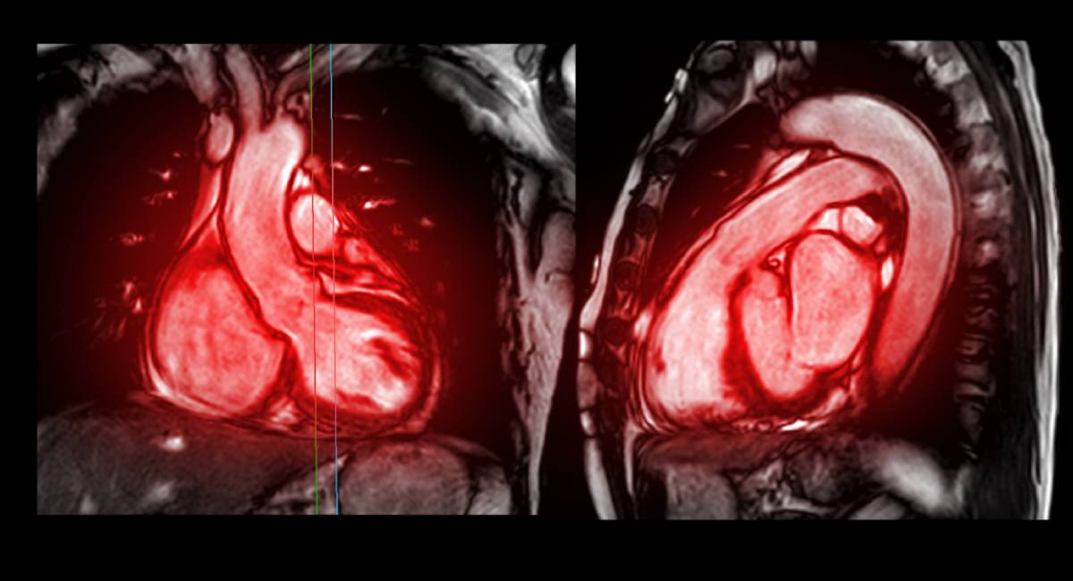
Cardiac MRI
Cardiac MRI uses magnetic fields and radio waves to produce high-resolution images. It is excellent for evaluating heart muscle, congenital disabilities, and complex conditions.
Nuclear Cardiac Imaging
This technique involves injecting a small amount of radioactive material to highlight areas of reduced blood flow. It is commonly used in stress tests to assess heart function under exertion.
When Should You Consider Cardiac Imaging?
You may need heart imaging if you experience symptoms like:
- Persistent chest pain or discomfort
- Unexplained shortness of breath
- Frequent dizziness or fainting spells
- Irregular heartbeats or palpitations
- Unusual fatigue during physical activity
Additionally, if you have risk factors such as high blood pressure, high cholesterol, diabetes, or a family history of heart disease, your doctor might recommend routine imaging.
Preparing for a Cardiac Imaging Test
Preparation varies depending on the type of imaging. Generally, you may be advised to:
- Avoid eating or drinking for a few hours before the test
- Wear comfortable clothing
- Inform your doctor of any medications you are taking
- Remove metal objects if undergoing an MRI
Always follow your healthcare provider’s instructions for the best results.
Is Cardiac Imaging Safe?
Most imaging tests are safe with minimal risks. Non-invasive procedures like echocardiograms and MRIs pose no radiation risk. CT scans and nuclear imaging involve low doses of radiation, but the benefits often outweigh the risks, especially when detecting serious heart conditions.
The Role of Cardiac Imaging in Preventive Care
Cardiac imaging is not just for diagnosing problems; it is a vital tool for preventive care. Identifying potential issues sooner rather than later allows for timely interventions, reducing the risk of heart attacks, strokes, and other complications. Regular screenings, as recommended by your doctor, can significantly improve heart health outcomes.
Cardiac Imaging in Los Angeles
Cardiac imaging is an invaluable resource in modern cardiology, offering detailed insights into heart health. Whether diagnosing conditions, monitoring treatments, or assessing risks, these tests play a crucial role in comprehensive cardiac care. If your doctor recommends cardiac imaging, it is a proactive step toward understanding and protecting your heart health. Take that step forward with one of our cardiologists today.
Leave a Reply